Technology at Drill Dental
3D Dental Imaging
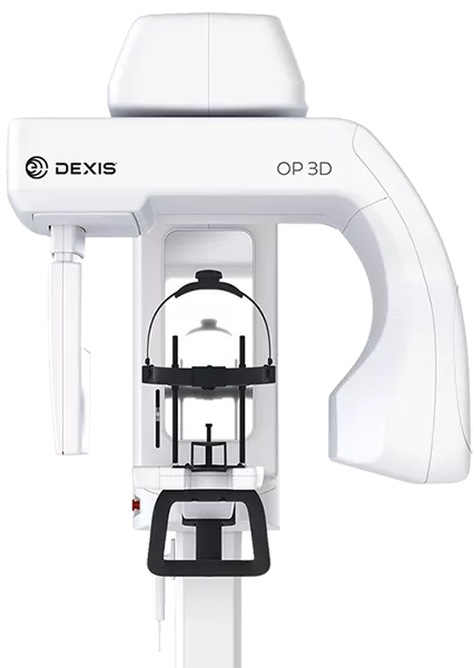
DEXIS OP 3D. Product image: DEXIS.
 Seeing beyond the surface
See more. Plan with a clearer picture. Our DEXIS CBCT scanner and DTX Studio software help us look inside the teeth and jaws, investigate concerns, and plan treatment in three dimensions.
Inside DTX Studio
A look at the views that help turn a scan into a clearer understanding of your care.
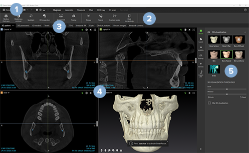
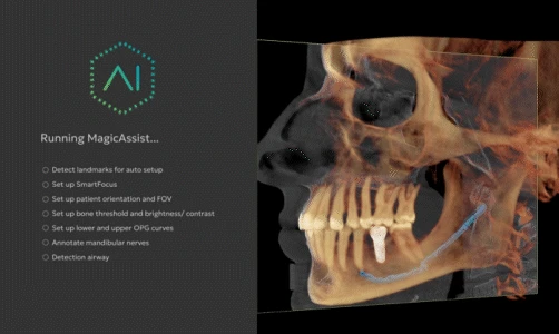
DTX Studio manufacturer demonstration screens. Images: DEXIS / DTX Studio. Diagnostic interface case courtesy of Dr. Katya Archambault. Tap either image to enlarge.
What a CBCT scan shows
A cone beam CT scan, or CBCT, uses X-rays to build a three-dimensional view of your teeth and jaws. In DTX Studio, we can turn that view and examine thin slices through it. This helps separate structures that overlap on a flat X-ray.
A closer look for diagnosis
When a specific concern needs more detail, CBCT can help us assess the shape of tooth roots, changes in the bone around a root, or the position of an impacted tooth. For selected root canal cases, it can help us investigate complex anatomy or a tooth that has not healed as expected. We connect the scan findings with your symptoms, examination, and other tests.
Planning before surgery
Before implant treatment, we assess the height and width of available bone and identify nearby structures, including nerve canals and the sinuses. A virtual implant plan helps us consider its size, angle, and position in relation to those structures and the tooth it will support. The scan also helps us assess whether bone grafting may be part of your plan.
Putting the digital picture together
DTX Studio can bring CBCT images and digital impressions together, connecting the bone beneath the gums with the shape of your teeth above them. We use these views to discuss the proposed treatment with you. When appropriate, digital planning can also support a surgical guide or another planned surgical approach.
What you can expect
For a CBCT scan, you are positioned in the scanner and asked to stay still as it moves around your head. The team explains the positioning before the scan. Your dentist will discuss what the images show and how the findings affect your options.
Questions patients ask
Does every appointment need a 3D scan?
No. Imaging is chosen to answer a specific clinical question. Your dentist decides whether an examination, existing images, conventional X-rays, or a 3D scan will provide the information needed.
Does a 3D scan use radiation?
Yes. CBCT uses X-rays and generally involves more radiation than a conventional dental X-ray. Your dentist recommends it when the additional information is appropriate for your care.